Testing CMFDA for Measuring Proliferation
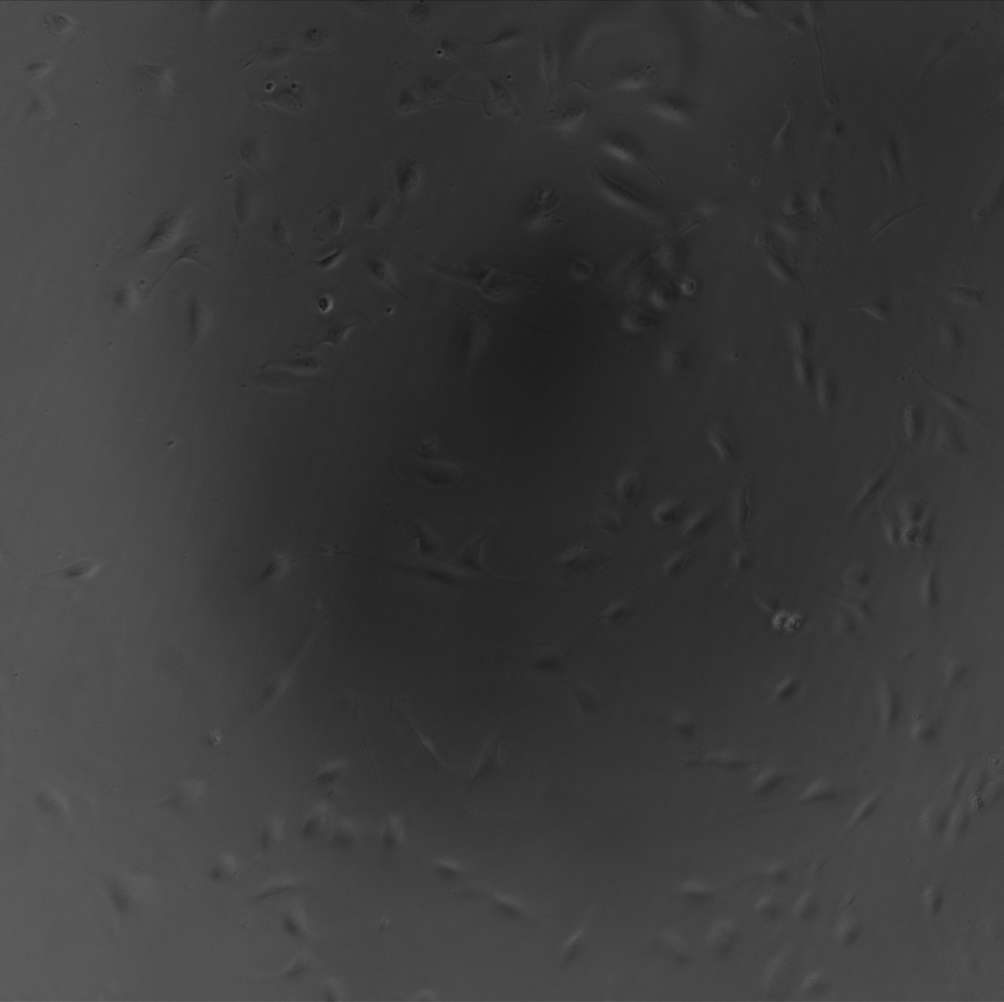
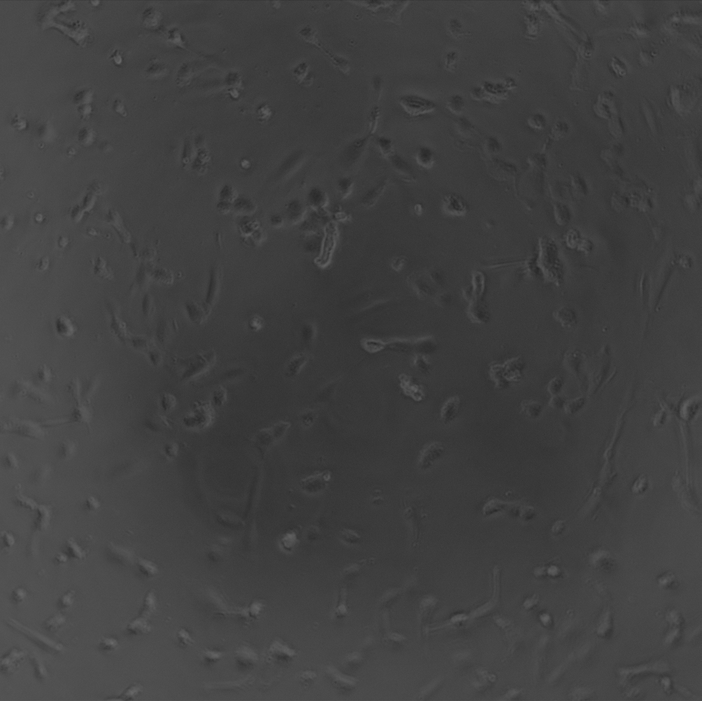
The goal of the experiment was to use a fluorescent dye to label live cells in order to measure proliferation over several days. To avoid cell toxicity, the intensity of the fluorescence was reduced to ~1.5%. Human umbilical vein endothelial cells (HUVECs) were seeded in a a 96-well plate at a density of 5000 cells per well. CMFDA, a live cell marker, was added at a concentration of 5uM on days 1 and 5. Images were taken at Days 1, 3, 5, and 8. Here are representative phase contrast images of Control (no CMFDA) and CMFDA (cells with CMFDA added and imaged at 1.5% fluorescent intensity):
Day 1
Control CMFDA
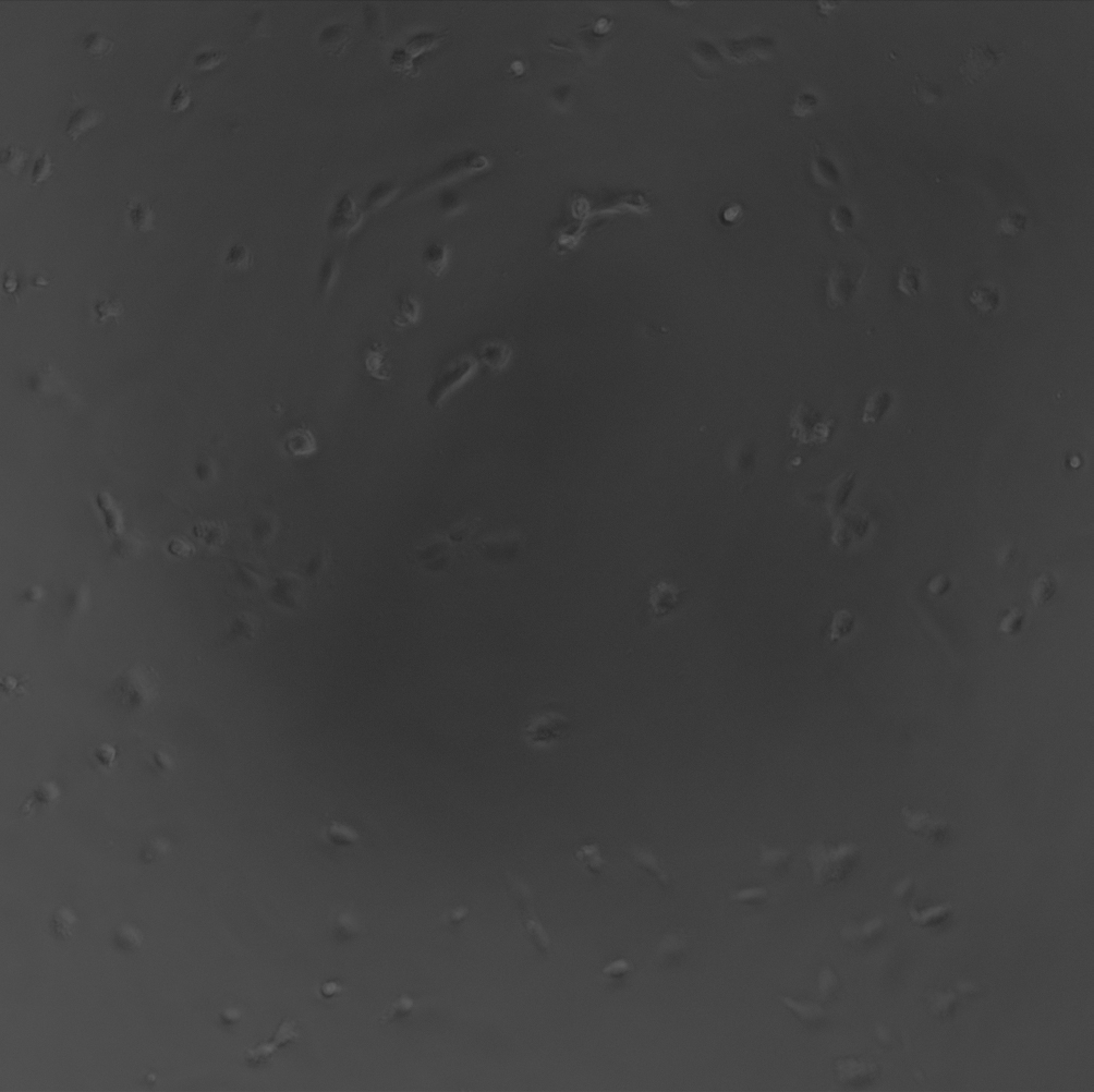
Day 3
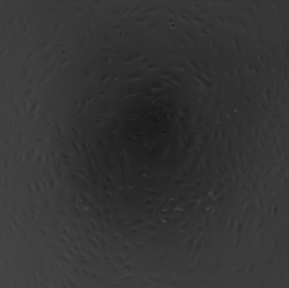

Day 5
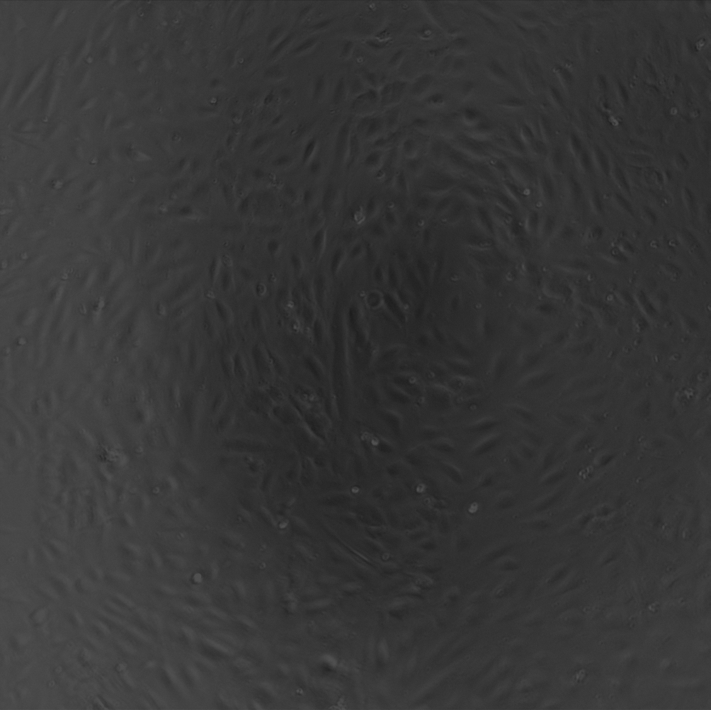
DAY 8
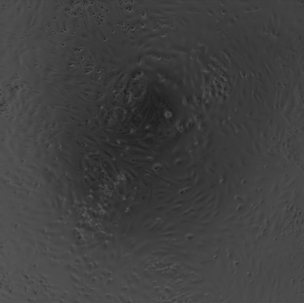

Stephanie counted the cells in ImageJ. In control wells, cells displayed a linear growth curve and became confluent by day 5. The days the CMFDA was added, days 1 and 5. Cells appeared rounded and apoptotic. On days 3 and 8, the cells seemed to recover and spread, but the CMFDA or fluorescent imaging affected cell viability/proliferation. At all time points, there was a significant decrease in cell number when CMFDA was added to the cultures.

We are planning on measuring proliferation on membranes of different pore sizes and porosity. Originally, we were going to stain with CMFDA, but our plans now are to take phase contrast images. Cells are distinguishable without fluorescence.