Filtration of Bioconjugated Samples Retains Gold, but We Can't Follow the Ligand
On Friday(which was a great day for science). I ran a series of 5 passes of the bioconjugated gold nanoparticles through 30nm cutoff Gen 3 Sepcons. If you’ll remember, the Barcikowski lab wants the free ligand separated out from the complexes.
To indirectly get the relative concentrations of gold and ligand, we measured the absorbance spectrum of the various samples. The Barcikowski lab had told us that the ligand absorbs at 280nm, and provided the following standard curve:

With a peptide concentration of 20uM (that of the pure sample) we would expect an extinction coefficient of 0.06 at 280nm. Since we know gold absorbs at around 515nm, the TECAN was made to scan from 230-700nm at intervals of 5nm. After some inconclusive results taken last week, I first ran scans of each window of the TECAN plate with just pure buffer: 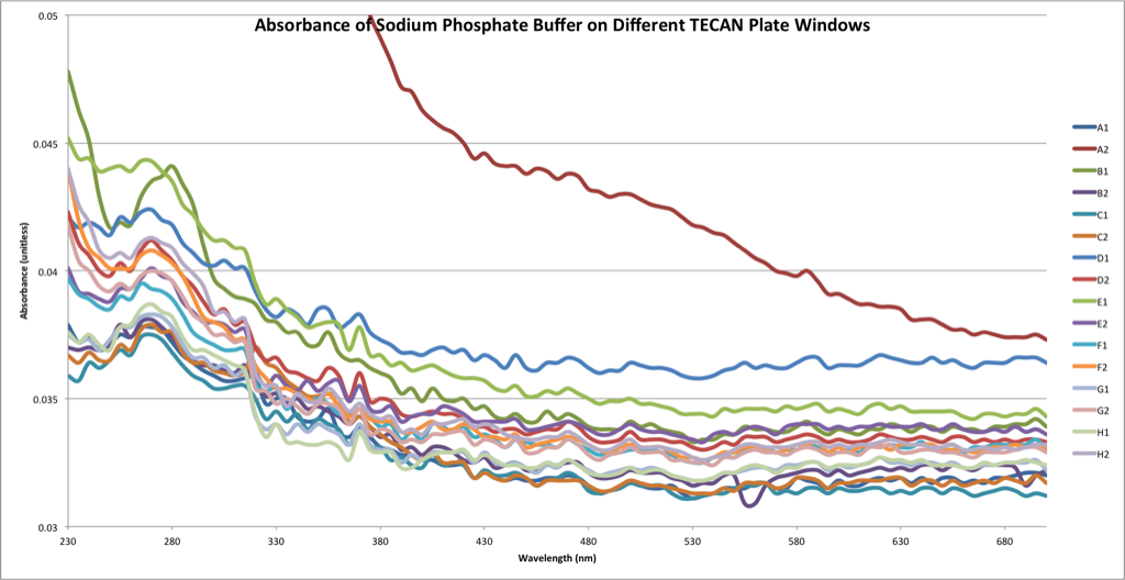
The purpose of this was two-fold. First, A1 (Auburn) and B1 (Forest Green) are sufficiently odd that I did not use them for any of the remaining data collection. Additionally, since the bands seem to differ by some constant, in the graphs that follow I simply subtracted the buffer signal from the appropriate window from the new signal. First, the final results:

The following represents the sequence of passes:

Which gave rise to the following progressive filtrates:

and the following retentates:

Two immediate points: First, the vagaries of the TECAN measurements could possibly be due to protein fouling on the surface of the plate I’m using. I don’t clean with an acid-base wash, but just ethanol. I’m hesitant to try acid-base, because the plate already looks etched, as though someone tried it once. I’ll be looking into this. EDIT – the manual recommends using ethanol to clean it. Secondly, why doesn’t ultracentrifuging the pure sample give us a peak in the 280nm range? I coated all of the centrifuge vials with 20uM albumin and washed thoroughly with buffer before adding the samples, so I don’t believe adsorption is the source of our troubles. Also, we’d thought that the strong signal for 1F might have been due to BSA contamination, but this doesn’t seem to be the case – below is the spectrum for pure BSA, which looks nothing like 1F’s peak-less signal:
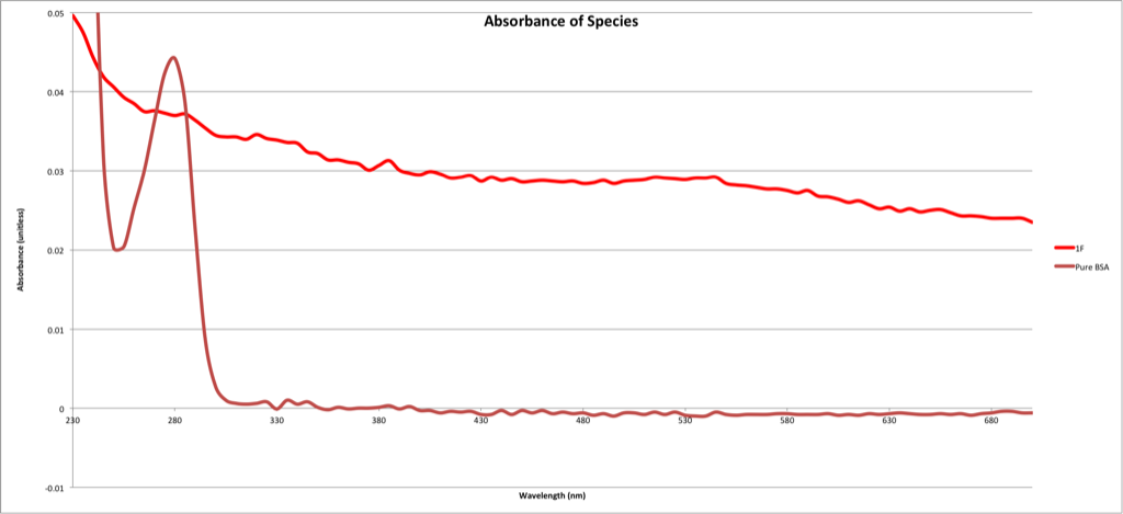
I looked at the paper the Barcikowski lab released about penetratin-conjugated gold nanoparticles (Penetratin-Conjugated Gold Nanoparticles), and they measure free ligand in the same way – ultracentrifugation at 30,000g for 30 minutes, then TECAN absorbance at 280. I’m not sure why we get no signal when they do, and in the hopes of resolving this I gave the Barcikowski lab a small update detailing this problem of the missing ligand.