MEM Elution Assays on pnc-Si Membranes – Rotation Summary
The minimum essential medium elution assay with neutral red is a commonly used method for determining the cytotoxicity of a material, typically polymers1-4. The neutral red dye is only endocytosed by viable cells; relative viability can be determined by absorbance measurements from solutions of the extracted dye. After several weeks in the lab stabilizing the cell line and conducting dry runs of the experiment, two elution assays were completed. The first assay examined effects after one day (24 hours) of exposure to the elution and the second assay examined effects after both one and five days of exposure. While dramatically toxic effects are not known to be associated with SiMPore pnc-Si membranes, inconsistencies within these results cannot allow for a robust conclusion.
L929 cells, murine tumor fibroblasts, are the cell of choice for this assay1,3, and were used for both experiments, at a passage number on the order of 570. They were cultured with Gibco’s MEM (10% BCS, 1% Pen-Strep, 1% non-essential amino acids). A 24 well plate was used for cellular incubation with elution, for both experiments. Negative control wells received 0.5ml of 0.9M NaOH while positive control wells received the same amount of fresh media. In each case, the neutral red solution was made at a concentration of 50 ug/ml, dissolved in media. The fixing solution was 4% formaldehyde, and the extraction solution was 1% acetic acid in 50% ethanol.
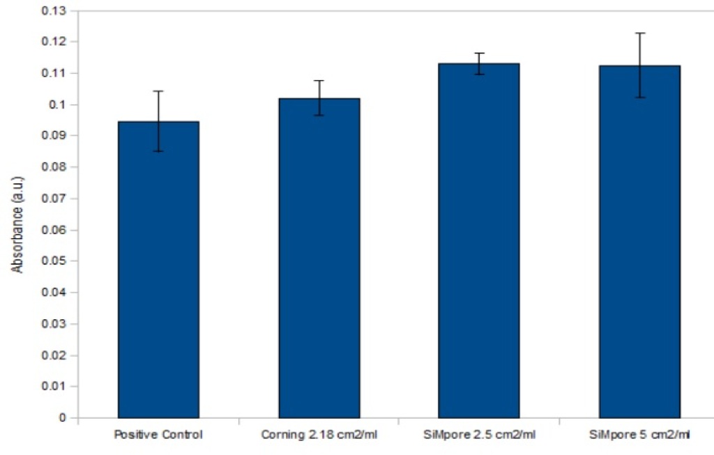
For the first experiment, five SiMPore pnc-Si culture and five Corning polyester insert membranes were incubated in media for 24 hours, with comparable surface area to volume ratios. At the same time, L929 cells were plated in the 24 well plate at 50,000 cells per well and allowed to settle for the same 24 hours. At the end of this period, 300ul of elution for each of the different SA:V ratios was transferred to the cell plate and incubated for an additional 24 hours. Negative and positive controls were introduced at this time. At the end of this incubation period, the elution in all wells was replaced with 0.5ml of neutral red solution, incubated for three hours then fixed for 20 minutes. After fixation the wells were washed with HBSS and the dye was extracted with agitation for 10 minutes. Absorbance data was taken with a TECAN spectrophotometer at 540nm and the background reading from wells containing only the extraction solution was subtracted before analysis. One way ANOVA was used to determine significant differences, with a Duncan post hoc analysis as well (p<0.05). Error bars show standard error; n=3 for each group. Significance was only found in comparison to the negative control (zero absorbance), as expected.
Results show no significant differences between any experimental groups and positive control, leading to a conclusion of minimal to zero toxicity of both membrane types. Very similar phase contrast images of the cells in each group support this conclusion, as previously shown during lab meeting.
The second experiment was performed in much the same manner, with significant contribution from Greg Madejski during all stages. However, the experimental groups examined here included two types of SiMPore membranes (C-300 and C-1000) in addition to the Corning membranes for comparison. After incubation of the membranes with media for five days, the Corning plate showed almost complete evaporation, which was both unexpected and inconvenient. To circumvent this, new Corning membranes were peeled off their plastic insert frame and sealed in a 15ml falcon tube with media for the five day period; this is a slightly different arrangement than was previously used, which was incubation in a six well plate. During this additional incubation period the day one and SiMPore day five elutions successfully obtained were refrigerated. The SiMPore day five elutions also experienced some evaporation, but only lost about 1ml.
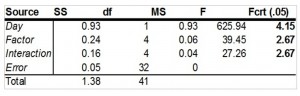
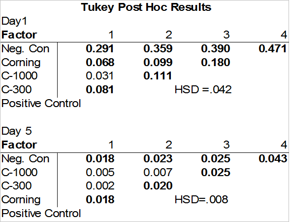
Once all elutions were obtained and brought up to 37C, they were transferred to one 24 well plate for day one and day five, 300ul elution per well. These plates were incubated for 24 hours with the elution, then processed as in the first experiment. A multifactor ANOVA was conducted on the data. A significant interaction effect was found between the days and the experimental groups, so individual Tukey HSD post hoc analyses were conducted for each day. Significant differences were found between all experimental groups and both controls; bolded values in the tables indicate significance.
While somewhat encouraging, these results may be ultimately unreliable based solely on the inconsistencies between positive control groups and variability introduced when dealing with such small absorbance values. In the first experiment the positive control mean absorbance was smaller than all experimental groups, which should not have been the case. In the second experiment the mean absorbance of the positive control drastically decreased from day one to day five; this also should not have been the case. These differences, in addition to the large error, inherently preclude concrete comparisons. After consideration of the protocol, whether or not the media that was incubated for five days was allowed to evaporate may have caused some change that is responsible. The decrease in the day five positive control as compared to day one indicates some difference exists outside the experimental factor has affected the data; at this point the cause of this difference is still unknown.
Figures:
1. Experiment One: Mean Absorbance Values. Experimental groups from left to right are positive control, Corning SA:V ratio of 2.18 cm2/ml, SiMPore SA:V ratio of 2.5 cm2/ml, SiMPore SA:V ratio of 5cm2/ml; n=3 for each group.
2. Experiment Two: Mean Absorbance Values. Orders of magnitude difference between day one and day five values; no trend. Experimental groups from left to right are positive control, SiMPore C-300, SiMPore C-1000, and Corning. Day one left to right, n=3, n=6, n=5, and n=4. Day five left to right, n=3, n=5, n=6, n=3.

3. Multifactor ANOVA results for Experiment Two. F values (bolded) were significant for day, factor, and interaction.
4. Post Hoc Analysis Results: Tukey Test. Bolded values indicate significance. The only difference not significant in the Day One data is between the SiMPore membrane groups. Day five data shows significant differences between the controls and all experimental groups.
References:
[1] Repetto, Guillermo, Ana Del Peso, and Jorge L. Zurita. “Neutral Red Uptake Assay for the Estimation of Cell Viability/cytotoxicity.” Nature Protocols 3.7 (2008): 1125-131. Print.
[2] Borenfreund, Ellen, and James A. Puerner. “A Simple Quantitative Procedure Using Monolayer Cultures for Cytotoxicity Assays (HTD/NR-90).” Journal of Tissue Culture Methods 9.1 (1985): 7-9. Print.
[3] Hexig, Bayar, Ryusuke Nakaoka, and Toshie Tsuchiya. “Safety Evaluation of Surgical Materials by Cytotoxicity Testing.” Journal of Artificial Organs 11.4 (2008): 204-11. Print.
[4] Sigusch, Bernd W., Torsten Pflaum, Andrea Völpel, Matthias Schinkel, and Klaus D. Jandt. “The Influence of Various Light Curing Units on the Cytotoxicity of Dental Adhesives.” Dental Materials 25 (2009): 1447-1452.