Blood brain barrier co-culture (trial 3)
It’s been a while since I’ve done long-term BBB co-culture experiments on transwells but I recently completed a study. I was particularly interested to determine whether I could reproduce this spike in TEER that I measured in 1 previous experiment. I also needed repeats anyway.
I followed the normal procedure – seeded P11 bEnd3 on the pnc-Si side, facing down, and P18 NG10815 on the well-side facing up. Both were at 50000 cells/cm2. I measured the TEER every other day until day 9 and then waited until day 14 for the last TEER. I fed the cells every other day and stained on day 14. For controls, I grew monocultures of these cell lines. I started out with 2 transwells (PET and pnc-Si) of each condition. Unfortunately, 1 of the bEnd only pnc-Si transwells broke after 1 day and 1 of the co-culture pnc-Si transwells broke as I measured it on day 14.
TEER data show no transient spike. Both co-cultures had a much higher increase in TEER than the bEnd samples which suggests that the glial cells are signaling to tighten the endothelial barrier. Note the increase in TEER and the final % increase) is faster and higher, respectively, on pnc-Si than PET. Also, the TEER was trending down by day 14, which may support my hypothesis that the rapidly acidified NG10815 media harms the endothelial cells on the other side of the membranes. The TEER of NG101815 samples increased more quickly on pnc-Si due to the familiar clumping of those cells in the wells of the chips. I would say the TEER of bEnd3 cells alone was ‘normal’.

Live/Dead stain of bEnd3 only and NG108-15 only. These cells exhibit normal morphology and viability.

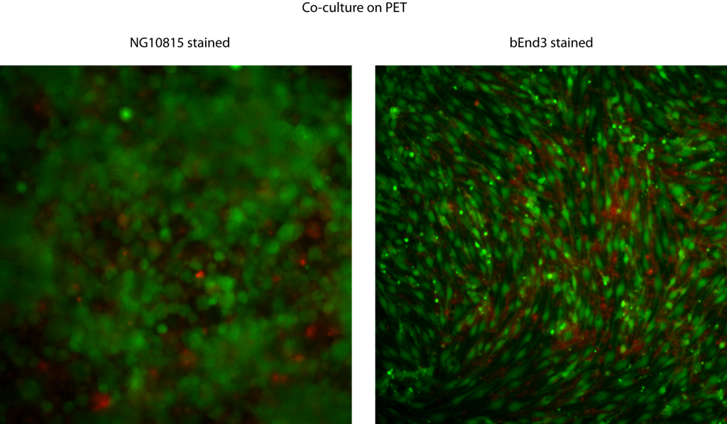
Live/Dead stain of cocultures on PET (top) and pnc-Si (bottom). For PET samples, I stained the NG10815 layer only (left) and the bEnd3 layer only (right). Both cell types look as they would if they were cultured by themselves.
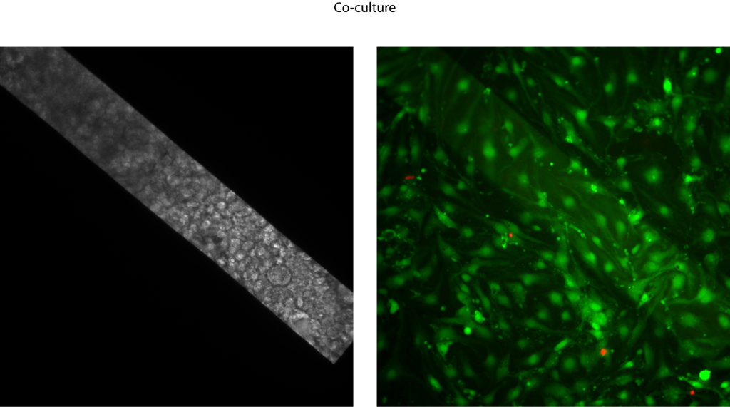
I was only able to get a picture of the bEnd3 component of the co-culture since I only had 1 sample to stain. The bEnd2 cells look weird – the density is not comparable to a monoculture, there are no vacuoles over the free-standing membrane and the morphology doesn’t look all that great. I’ve noticed the lack of vacuoles on other BBB co-cultures (here and here), although not with every sample. Could it be that in those samples where the NG10815 cells totally block the well-side, they block permeability and the bEnd3 cells no longer express vacuoles? So, the combination of weird bEnd3 morphology with the lack of a spike in the TEER measurements makes me a little puzzled. However, the TEER data does support my original hypothesis of synergistic barrier function with co-cultures.
UPDATE(MAY 17, 2010): TEER Data as “Net TEER”
