Endothelial actin networks on pnc-Si transwells
I’ve been wondering if there were differences in actin/cytoskeletal structure of cells on supported vs. free-standing pnc-Si. A couple weeks ago, I had a couple of extra pnc-Si transwells so I stained for actin.
I followed the actin staining (rhodamine-phalloidin) method I uploaded to the protocols section of the lab, and I counterstained the nuclei blue with Hoechst 33342. The bEnd3 cells were grown on SC500 transwells for 14 days before staining. I used the 20X objective on the Zeiss for these images.
Commercial PET membrane:
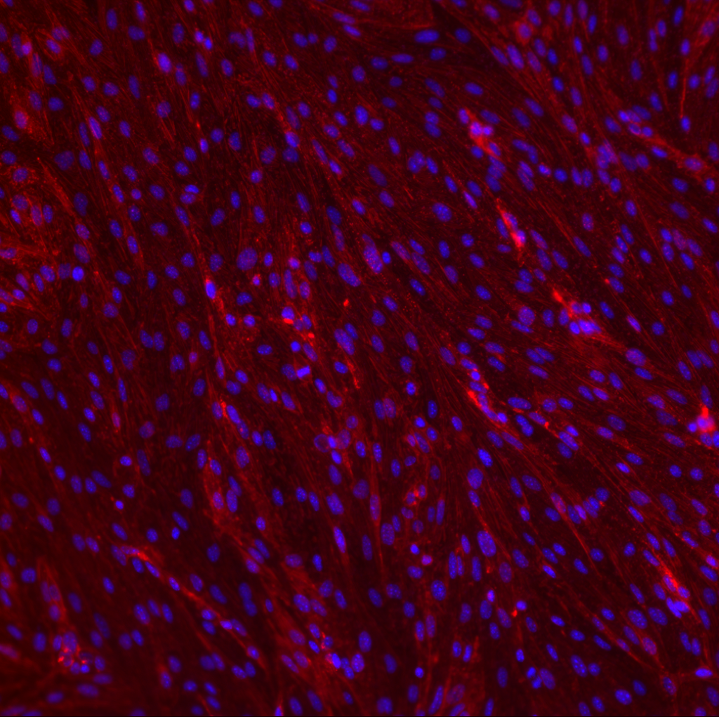
This image shows faint staining of actin filaments that are generally aligned with cell nuclei.
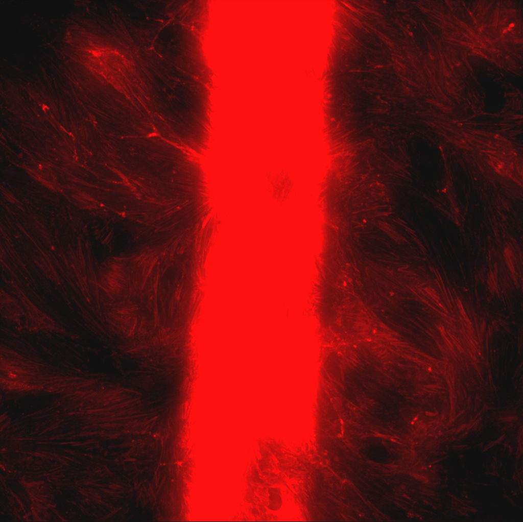
DIC (top), nuclei (bottom left) and actin (bottom right) of cells on SC500 transwell:
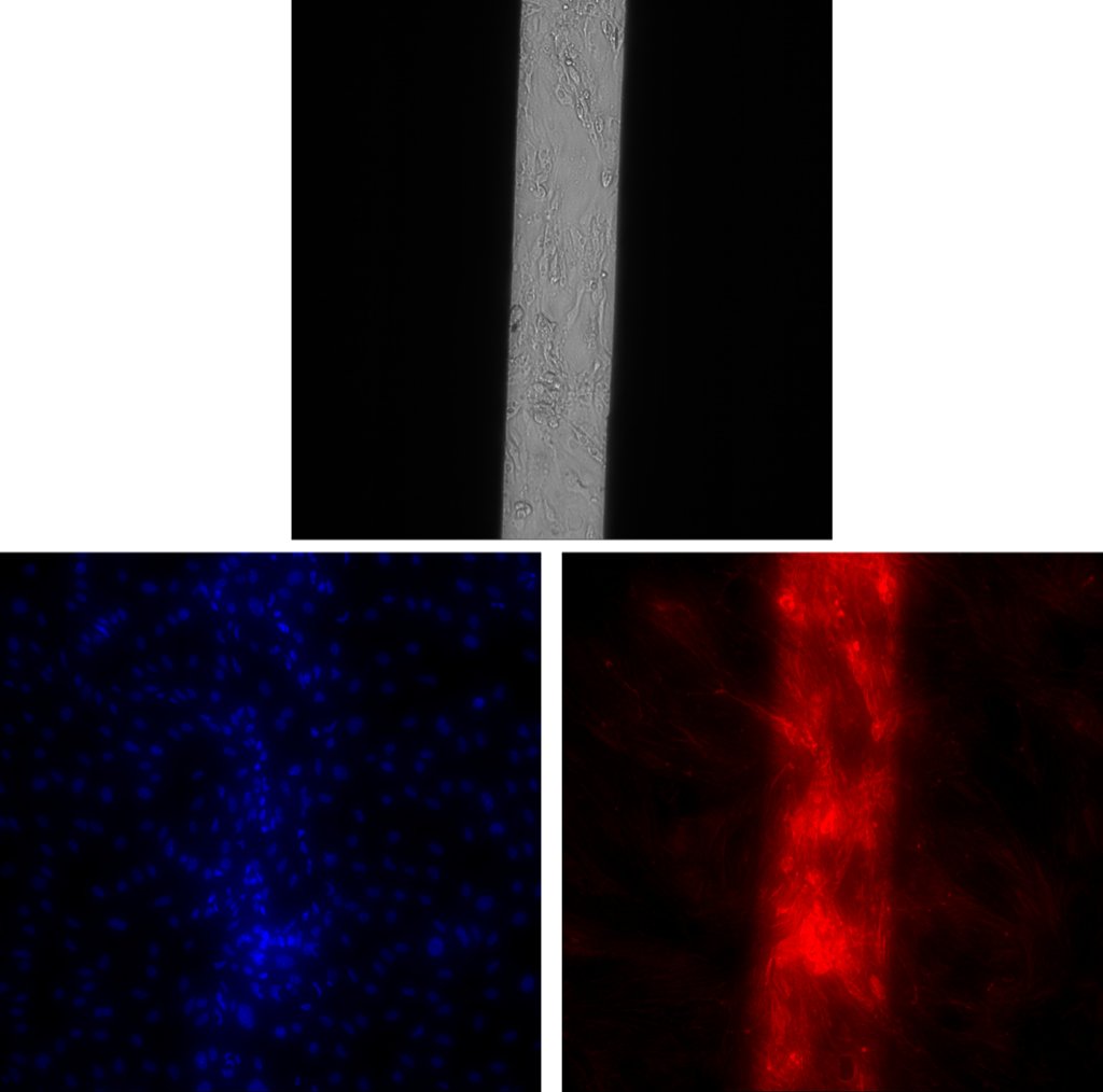
In DIC, you can clearly see many vacuoles. In both blue and red channels, the fluorescence was much, much brighter over the membrane window than over the supported pnc-Si. I see this phenomenon with green calcein AM, also. These cells were fixed before I stained them, so the enhanced fluorescence can’t be due to more active endocytosis/metabolism of cells on the window. Thus, this result supports the idea that supported pnc-Si (or the Si wafer) captures some of the emitted fluorescence. Interestingly, cell density on free-standing pnc-Si appears to be higher than on supported pnc-Si (based on counting the nuclei). Also, the nuclei appear more elongated over the membrane window. I brightened the red channel to show cells on the supported pnc-Si (below). The actin filaments seem to be more randomly aligned on supported pnc-Si than over the membrane window. However, I’m not sure if this is an effect of membrane mechanics/porosity or cells crowding onto the membrane. Based on these images, I don’t think actin staining will help elucidate any of our free-standing vs. supported pnc-Si cell questions. Also, I think the images would be more illustrative at 100X, which requires more extensive sample prep with Sepcons.