Endothelial cells form vacuoles within 1 day on pnc-Si
In this experiment, I wanted to test how quickly vacuoles form in endothelial cells when cultured on pnc-Si transwells. Here I used 30nm SC502 RTP’ed samples with the pnc-Si side down. I seeded P9 bEnd3 cells at 50000 cells/cm2 on the pnc-Si side, allowed the cells to attach for 2 hours and then inverted the transwells in a 24-well plate. I then used Live/Dead solution to stain the cells 1 (and 4) days later and imaged at 20X.
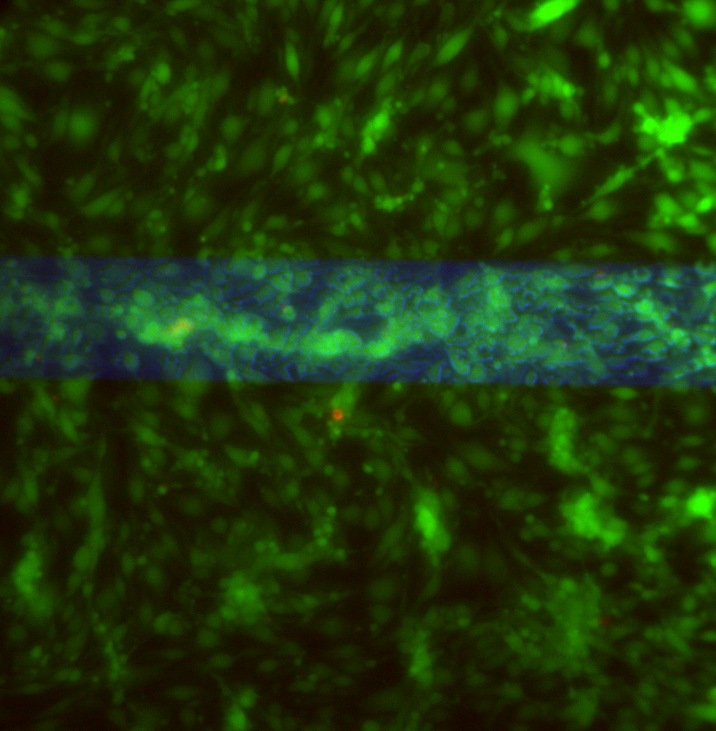
The phase image isn’t loading correctly, so here are 2 different areas on 1 sample (live=green, red=dead, blue=transmitted light). The top image is much better because I removed the sample from the 24-well plate and imaged through a coverslip. You can clearly see vacuoles in the cells only over the free-standing membrane.

I also stained a sample at 4 days post-seeding:


In these day 4 images, vacuoles are still present over the membrane and you can even see them in phase contrast.
These results show that the endothelial cells have formed vacuoles within a day of seeding on pnc-Si membrane transwells.
This is good news because it means you can make assessments of the conditions that cause the phenomenon pretty quickly. Are you sure you can’t see them in phase at 1-day?
Might be able to see vacuoles in phase at day 1 but the cell morphology was weird and the density was so high that it was difficult. I can check in the next experiment.