BMES 2018 Takeaways
BMES – Atlanta 10/17/18-10/20/18
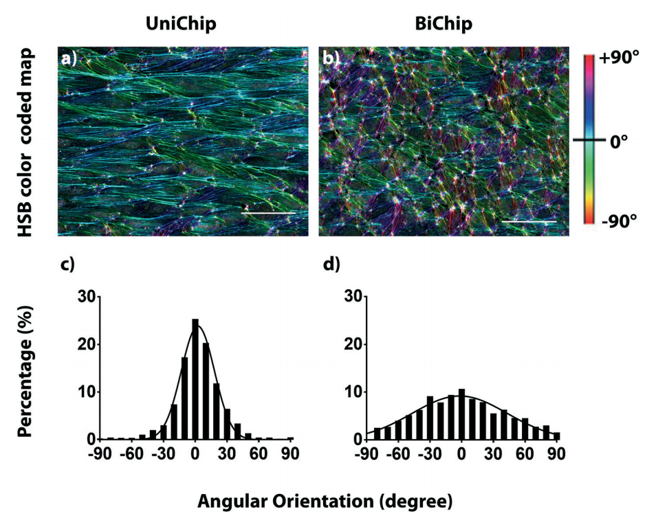
F-actin Alignment Quantification Using HSB Color Coded Maps
ImageJ Plugin: OrientationJ
Purpose: Quantification of F-actin alignment using ICC. HSB map provides a visual representation of fiber alignment with color codes related to hue:local orientation, saturation:coherency, brightness:fiber stain intensity (Figure 1).
Our use: Quantification of HUVEC alignment before and after flow.
Fluorescent TNF-alpha for Time Resolved Diffusion
Megan Catterton’s group presented their work on local stiumation of lymph node slices in a microfluidic device. The mentioned local stimulation of the lymph node using fluorescent TNF-alpha to watch the diffusion into the tissue slice. I would like to use this molecule to observe diffusion of my TNF-alpha from the luminal compartment to the abluminal compartment in my polarized signaling experiments. Further, I could use this to observe residual TNF-alpha after wash. (I found a reference but was unable to find a vendor for fluorescent TNF-alpha…)
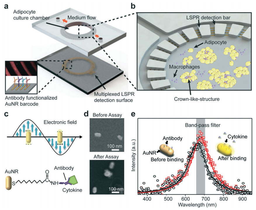
Ring Resonator on a Chip
Weiqiang Chen’s group presented their biosensor on a chip for the detection of adipose derived cytokines. While this is not my area of research, I believe it could be a nice direction for the biosensor on NPN project (if interest is still there). Reference: https://www.ncbi.nlm.nih.gov/pubmed/30302487
Abrupt Transitions in Matrix Stiffness Create Endothelial Stress Concentrations
Reinhart-King’s group presented on the use of PDMS posts to mimic local changes in vascular wall stiffness leading to abnormal F-actin and focal adhesions. I believe a major shortcoming of this study was the use of focal adhesion staining (vinculin on confluent cells…) on a non-continuous substrate. This could be an interesting direction for Tom’s group to take some of the micropatterning work; Hypothesis: Discrete microscale patterning regulates focal adhesion formation independent of matrix stiffness. This was shown before with Stephanie’s work, but this could be a good way to reach a greater audience with your papers to come.
Neutrophil Dyanmics
Bahareh Behkam’s group presented a talk titled ‘A Microfluidic Assay for High Resolution Quantitation of Dynamic Neutrophil Responses to Pathogens’. They had a simple PDMS device with two channels (side by side). They added neutrophils to one side and bacteria to the other. I believe this work was very lackluster in execution, but interesting in theory. We could definitely improve on this by using our actually physiological set up (not just glass) to observe neutrophil dynamics in response to bacteria. One thing I did like out of the talk was their interest in neutrophil directional persistence (DP), where DP = displacement / distance traveled. This results in a number between 0 and 1 that quantifies the effective persistence of the neutrophil in movement. I think this could be a really interesting value when observing differences in fMLP and TNFa mediated neutrophil activation.
General Thoughts
Breast cancer cell metastasis is a really popular biomedical engineering area of interest right now. I believe with little modification we could answer some interesting question on how these cells migrate and what they migrate to in our NPN flow device with HUVECs. I didn’t attend any talks that I believe improved on our platform design in this area of interest or in the neutrophil dynamics area.