hiPSC Differentiation to Brain Microvascular Endothelial Cells (BMEC)
INTRODUCTION
The blood-brain barrier (BBB) is a highly selective endothelial barrier that serves a crucial role in maintaining tissue homeostasis in the brain. Many current models, however, suffer from low TEER and permeability values far greater than physiological levels. Recently, human induced pluripotent stem cells (hiPSC) have been differentiated into BMECS with very high TEER and good expression of tight junction proteins. We sought to replicate this protocol in our lab with non-patient specific iPSCs in order to develop a BBB model in our microchip devices.
METHODS
Differentiation
Before differentiation (D-3):
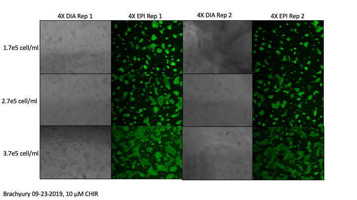
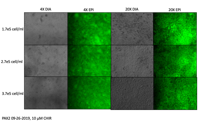
hiPSCs (WiCell, [iPSC(IMR90)-4]) were singularized with Accutase and plated onto the vitronectin-coated plates at a surface density of 1.7 x 10^5, 2.7 x 10^5, or 3.7 x 10^5 cells/mL (equivalent to 3.7 x 10^4, 5.8 x 10^4, and 7.9 x 10^4 cells/cm^2 based on email from Britta’s employee) in mTeSR1 supplemented with 10 µM ROCK inhibitor Y-27632 in a 24-well plate. hPSCs were expanded in mTeSR1 (no ROCK inhibitor) for 3 days.
Experiment 1: A separate set of cells were plated at 50 x 10^3 cells/cm^2 and expanded as described above and processed for immunochemistry to check for markers of pluripotency, Oct-4 and Nanog (Day 0 of differentiation, parallel cells).
To initiate differentiation at day 0 (D0):
Experiment 2: Cells were treated with 0, 6, 8, and 10 µM CHIR99021 in DeSR1 (DMEM/Ham’s F12, 1xMEM-NEAA, 0.5xGlutaMAX, and 0.1 mM beta-mercaptoethanol).
Experiment 3: Cells were treated with 10 µM CHIR99021 in DeSR1 (DMEM/Ham’s F12, 1xMEM-NEAA, 0.5xGlutaMAX, and 0.1 mM beta-mercaptoethanol) and maintained in this medium for 4 days.
Primitive Streak (D1):
The medium was changed to DeSR2: DeSR1 plus 1x B27 every day for another 4 days.
Experiment 2: Cells were processed for immunochemistry to check for a primitive streak marker, Brachyury.
Experiment 3: A separate set of cells were treated as described above and processed for immunochemistry to check for a primitive streak marker, Brachyury.
Mesodermal Intermediate (D4):
Experiment 3: A separate set of cells were treated as described above and processed for immunochemistry to check for a mesodermal intermediate marker, PAX2.
Endothelial Cell Progenitors (D5):
Experiment 3: A separate set of cells were treated as described above and processed for immunochemistry to check for an endothelial cell progenitor marker, VEGFR2.
To drive BMEC specification (D6):
The medium was changed to hECSR1 (hESFM, 20 ng bFGF, 10mM RA, and B27) for 2 days.
Replate cells in 96 well plate, Transwells, and devices (D8):
Cells were singularized with Accutase and plated onto Collagen IV, Fibronectin-coated plates at a surface density of 1 x 10^6 cells/cm^2 in hECSR1 and maintained in this medium for one day.
BMEC maintenance (D9):
The medium was changed to hECSR2: hECSR1 lacking bFGF or RA for one day (multiple days for Transwells).
BMEC characterization (D10):
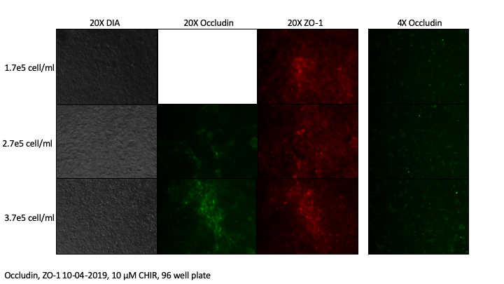
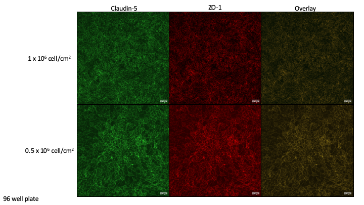
Cells were processed for immunochemistry to check for tight junction markers, ZO-1, Occludin, and Claudin-5.
Immunochemistry
Cells were rinsed with ice-cold phosphate-buffered saline (PBS) once, fixed in 4% paraformaldehyde (PFA) for 15 min. Cells were then blocked with 10% goat serum in PBS containing 0.3% Triton X-100 for 30 min (10% PBSGT). Modifications to this protocol were made for Brachyury staining and ZO-1/Occludin/Claudin staining based on the published methods paper.
Primary antibodies:
Antibodies were diluted in 10% PBSGT (except Brachyury), and cells were incubated in the antibody solutions at 4°C overnight.
Secondary antibodies:
After three PBS washes, cells were incubated with secondary antibodies in 10% PBSGT (goat anti-mouse Alexa Fluor 488, except Brachyury and ZO-1) for 1 hour at room temperature. Cells were then washed with PBS three times and were visualized.
RESULTS
Immunochemistry
D0 Parallel, Markers of Pluripotency

Experiment 2: D1, Marker of Primitive Streak (optimizing CHIR and seeding densities)

Experiment 3: D1, Marker of Primitive Streak (optimizing seeding densities)

Experiment 3: D4, Marker of Mesodermal Intermediate (optimizing seeding densities)
Experiment 3: D5, Marker of Endothelial Cell Progenitors (optimizing seeding densities)


Experiment 3: D10, Tight Junction Markers to characterize BMECs (optimizing seeding densities)
96 Well Plate Staining:

Microchip Device Staining:


I did not include images from other devices and seeding densities, but the results were comparable to what we saw in the 96 well plate.
TEER
Need new EndOhm cup. Chopstick method was done quickly (had to run to class), with measurements same as background. Unclear if this is due to gaps in monolayer, poor pipetting, poor acquisition, etc… Fixed cells and will check monolayer using cresyl violet.
DISCUSSION
We have successfully cultured hiPSCs, with moderately efficient differentiation into endothelial progenitor cells. We are currently expanding these cells to freeze and try differentiation again in hopes to save time in future experiments. It was necessary to optimize CHIR concentrations for our lot, increasing the published 6 µM to 10 µM. We had poor differentiation to endothelial progenitors our first time through (not included in post), which we believe was due to suboptimal active concentrations of CHIR. In addition, we found comparable staining at each check point, but different morphologies, for cells seeded at 1.7, 2.7 and 3.7 x 10^5 cells/ml. In our final product BMECs, while we could successfully grow cells on both platforms, staining was very heterogenous in both devices and 96 well plates, likely due to suboptimal differentiation efficiency.
Future Plans:
We will try seeding endothelial progenitors at different concentrations, as we appeared to have multiple layers of cells instead of monolayer, and will likely try cell sorting the endothelial progenitors prior to reseeding. We will optimize TEER protocols using hCMEC/D3s, purchasing a new EndOhm chamber to improve accuracy of reading, before measuring TEER on our hiPSC-derived BMECs.
UPDATE
METHODS (1)
Differentiation (Changes only)
Replate cells in 96 well plate, Transwells, and devices (D8):
Cells were singularized with Accutase (NEW METHOD: sat in Accutase 30 min+ minutes until dissociated and in single cell) and plated onto Collagen IV, Fibronectin-coated plates at varying surface densities: 1 x 10^6 cells/cm^2, 0.75 x 10^6 cells/cm^2, 0.5 x 10^6 cells/cm^2, or 0.25 x 10^6 cells/cm^2, in hECSR1 and maintained in this medium for one day.
RESULTS
Immunochemistry
D10, Tight Junction Markers to characterize BMECs (optimizing reseeding method and densities)
24 Well Plate Staining:

96 Well Plate Staining:
METHODS (2)
Differentiation (Changes only)
Replate cells in Transwells (D8):
Cells were plated onto Matrigel-coated 12-well and 24-well Transwell plates in hECSR1 and maintained in this medium for 4 days. TEER was taken each day using both EndOhm Chamber and STX methods.
RESULTS
TEER peaked around 20 ohm-cm^2 at Day 10.
DISCUSSION
In the first update experiment, we saw great junctional staining and monolayer formation. In the following experiment, cell morphology prior to replating was unusual, which may explain low TEER. TEER trends are the same (peak at D10) and size of Transwell does not appear to matter when replating.
PROTOCOL IS CHANGING. Our collaborators have highlighted issues with this protocol, and cells appear to be more epithelial than endothelial. They have been working on a novel protocol that is in final stages before submitting for publication. The cells they get with this protocol appear to be more endothelial and are representative of post-capillary venules (lower TEER, site of transmigration). I will put iPSC work on pause until I am trained on the novel protocol in Bern.