Cell Proliferation on Different Membrane Porosities and Pore Sizes
To determine whether cell proliferation is a function of membrane porosity and/or pore size, human umbilical vein endothelial cells (HUVECs) were cultured on various surfaces. The cells were seeded on low and high porosity membranes of 0.5 um and 3.0 um pore sizes. Non-porous membranes served as control membranes. All membranes were coated with Geltrex prior to cell seeding. As controls, the lower gasket was attached to the bottom of a 24-well plate, and cells were cultured on tissue culture plastic with and without Geltrex coating. In all conditions, 500 cells were seeded. Phase contrast images were taken at Days 1, 2 and 3. Images were then analyzed on ImageJ to count the number of cells. On porous membranes, only cells on the porous region were counted. To account for differences in seeding density, each well was normalized to its Day 1 cell number.
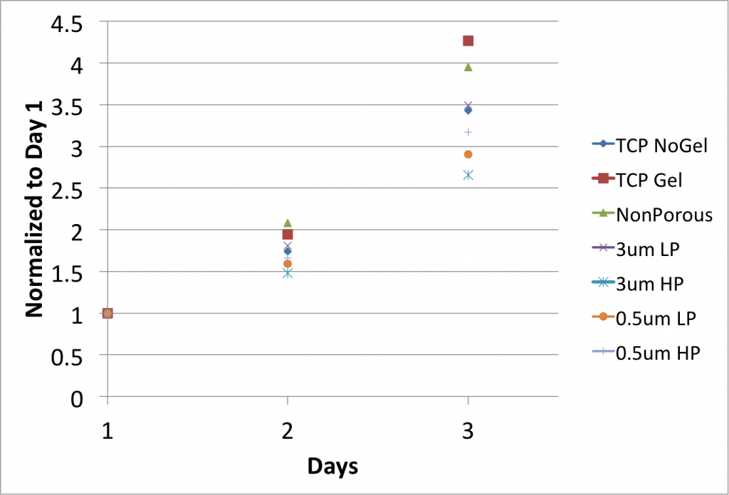
Because the sample size is n=2, statistical tests were not performed. However, it appears as if pore size and/or porosity affects cell proliferation. According to Andrea’s cell spreading data, HUVECs spread the most on TCP and the least on 3.0um pore size high porosity membranes. HUVECs displayed the highest proliferation on Geltrex-coated TCP and the lowest on 3.0um pore size high porosity membranes.
Cell spreading is correlated with proliferation. Restricting the spread area of adherent cells leads to growth arrest (Mih et al, 2012). The results suggest that by restricting cell spreading on porous membranes proliferation can be regulated. The experiment will be repeated to increase the sample size.