HUVEC 'proliferation' in Sepcons
Last week when I reported no membrane breakage after a week of HUVEC culture, I showed a phase micrograph on day 7. This week I tracked the HUVECs a bit more closely with the microscope.
This is 10X, HUVEC P5, initial seeding density of 32,500 cells/insert. I tried to capture approximately the same area on each day.
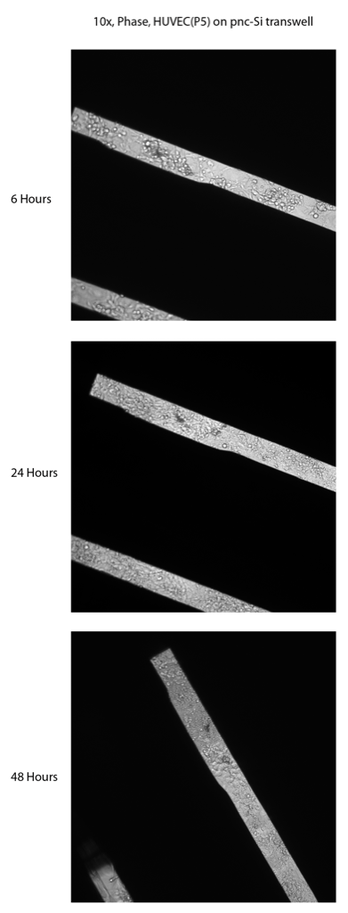
After 6 hours, the HUVECs are largely attached to the pnc-Si. You can see some HUVEC-looking cells in the background and clumps of cells in the foreground.
After 1 day in vitro, it seems like the clumpy looking cells have differentiated into a flatter phenotype. It seems like the membrane window is nearly covered by the cells. I can’t see a difference between the 24 and 48 hour time points (except that the bottom membrane broke). There appears to be at least 2 layers of cells because only certain cells are in focus. This is not ideal since we are hoping for monolayers. I think my seeding density might be too high, thus allowing cells to settle on top of each other. I’ll try lower seeding densities next week.
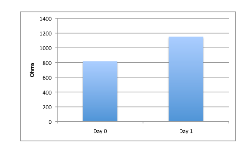
This increase in pnc-Si slit coverage by HUVECs was supported by Endohm data, which showed an almost 50% increase in TEER from day 0 to day 1.